Ultrasound in PCOS: What to Expect & What the Results Really Mean
If you’re on a journey to understand Polycystic Ovarian Syndrome (PCOS), you’ve likely heard that an ultrasound is a key part of the diagnosis. But what exactly are doctors looking for? What does it feel like? And what does it mean if they find “cysts” on your ovaries?
This guide will walk you through everything you need to know about the pelvic ultrasound in the context of PCOS, demystifying the process and the results.
First, a Quick Refresher: What is PCOS?
PCOS is a common hormonal condition that affects how the ovaries work. It’s characterized by a combination of symptoms, and no two people experience it exactly the same way. The widely used Rotterdam Criteria states that a diagnosis requires at least two of the following three features:
-
Irregular or Absent Periods: A sign that ovulation (the release of an egg) isn’t happening regularly.
-
High Levels of Androgens: These are “male” hormones (like testosterone) that can cause physical signs such as excess facial or body hair (hirsutism), acne, or hair thinning.
-
Polycystic Ovaries on Ultrasound: This is where the ultrasound comes in, and it’s often the most misunderstood part.
It’s crucial to know that you can have PCOS without having polycystic ovaries on an ultrasound, and you can have polycystic ovaries without having PCOS. The ultrasound is just one piece of the puzzle.
Why is an Ultrasound Used for PCOS Diagnosis?
The pelvic ultrasound gives doctors a live, inside view of your reproductive organs—specifically your uterus and ovaries. Its main jobs in the PCOS diagnostic process are to:
-
Check the appearance of the ovaries.
-
Measure the size and volume of the ovaries.
-
Count the number of small follicles present on each ovary.
-
Rule out other conditions that could be causing your symptoms, such as ovarian cysts or issues with the uterus.
What Are They Actually Looking For? The “Polycystic” Morphology
The term “polycystic ovary” can be scary because the word “cyst” often has negative connotations. However, in PCOS, these are not the large, problematic cysts that can cause pain and require surgery.
Instead, a polycystic ovary appears as an ovary with an abundance of very small, harmless follicles. Here’s what the sonographer and doctor are assessing:
-
Follicle Count: A key diagnostic feature. In PCOS, each ovary may contain 12 or more tiny follicles (ranging from 2 to 9 mm in diameter). These follicles are immature egg sacs that have developed but stalled due to the hormonal imbalances of PCOS, failing to release an egg.
-
Ovarian Volume: The ovaries in PCOS are often larger than average. An increased ovarian volume (typically greater than 10 mL) is another common characteristic. The high number of follicles causes the ovary to swell in size.
-
The “String of Pearls” Sign: This is a descriptive term you might hear. On the ultrasound screen, the many small follicles can look like a string of pearls lining the outer edge of the ovary.
In short: They are looking for ovaries that are larger than normal and packed with a high number of small, dormant follicles.
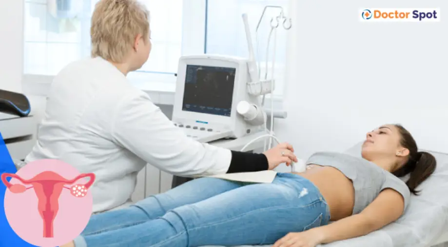
What to Expect During Your Pelvic Ultrasound
Knowing what will happen can ease a lot of anxiety. There are two main types of pelvic ultrasounds used, and sometimes both are performed for the clearest picture.
1. Transabdominal Ultrasound
-
How it works: A technician moves a transducer (a smooth, handheld device) over your lower abdomen.
-
Preparation: You will likely be asked to drink several glasses of water before the appointment and not go to the bathroom. A full bladder acts as a window, pushing gas-filled intestines out of the way to create a clearer image of your pelvic organs.
-
What it feels like: It’s generally painless, though there might be some mild pressure from the transducer, especially if your bladder is very full.
2. Transvaginal Ultrasound
-
How it works: A long, thin, sterilized transducer (about the width of a tampon) is gently inserted into the vagina. This allows the transducer to get much closer to the ovaries and uterus.
-
Preparation: You will be asked to empty your bladder right before the procedure.
-
What it feels like: It may feel a bit unusual or cause minor discomfort, but it should not be painful. You can always ask the technician to stop. The procedure is private and performed by a trained professional who will talk you through each step. A cover is always used on the transducer for hygiene.
Which one will you have? For adolescents or those who have never been sexually active, only a transabdominal ultrasound is performed. For most adults, a transabdominal scan is done first, often followed by a transvaginal one to get the most detailed view.
Understanding Your Ultrasound Results
After the scan, a radiologist will interpret the images and send a report to your doctor. Your doctor will then discuss the findings with you in the context of your other symptoms and blood test results.
Possible findings include:
-
“Consistent with PCOS”: This means your ovaries showed the classic signs of being polycystic (high follicle count and/or increased volume).
-
“Normal Appearing Ovaries”: This means your ovaries did not meet the criteria for being polycystic. Remember, this does not automatically rule out a PCOS diagnosis. If you have irregular periods and high androgens, you can still be diagnosed with PCOS.
-
Other Findings: The ultrasound might reveal a different issue, like a large functional cyst, fibroids, or other anatomical variations.
Limitations and Important Considerations
An ultrasound is a powerful tool, but it’s not perfect.
-
It Can’t See Hormones: The ultrasound can show the result of hormonal imbalances (the follicles), but it cannot measure your hormone levels. This is why blood tests are essential.
-
The “Right Time” Matters: If you are on hormonal birth control (the pill, IUD, etc.), it can change the appearance of your ovaries and mask the polycystic morphology. Your doctor needs to know your medication history.
-
Not a Standalone Test: A PCOS diagnosis is a clinical one, based on a combination of factors. The ultrasound is a crucial piece of evidence, but not the entire case.
Frequently Asked Questions (FAQs)
Q1: I have polycystic ovaries on my ultrasound, but my periods are regular. Do I have PCOS?
Not necessarily. Many people with no symptoms of PCOS can have polycystic-appearing ovaries. Without a second diagnostic feature (like irregular cycles or high androgens), you would not be diagnosed with PCOS.
Q2: Can an ultrasound tell me how severe my PCOS is?
No. The ultrasound shows the physical structure of your ovaries, but it doesn’t correlate with the severity of your symptoms. Someone with a very high follicle count might have mild symptoms, while someone with a lower count might have more severe ones.
Q3: Will I need more ultrasounds after I’m diagnosed?
It depends on your treatment plan and symptoms. If you are struggling with infertility and undergoing treatment, you may have frequent monitoring ultrasounds to track follicle growth. Otherwise, you likely won’t need another one unless new symptoms arise.
The Bottom Line
The pelvic ultrasound is a vital, non-invasive step in the PCOS diagnosis journey. It provides a visual clue to the hormonal chaos happening inside the body. But it’s just that—a clue.
Key Takeaways:
-
The “cysts” in PCOS are harmless, immature follicles, not large ovarian cysts.
-
The ultrasound looks for a high follicle count and enlarged ovarian volume.
-
A normal ultrasound does not rule out PCOS.
-
The results must always be interpreted by your doctor alongside your menstrual history and blood work.
Understanding what the ultrasound is and isn’t can empower you to have more informed conversations with your healthcare provider and actively participate in managing your health.
Disclaimer: This blog post is for informational purposes only and does not constitute medical advice. The diagnosis and management of PCOS should always be done in consultation with a qualified healthcare professional.